
Xray Glasses Archives Advanced Intelligence
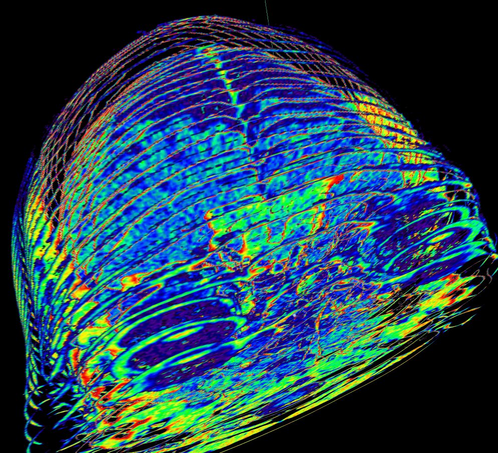
Stunning new images pave the way to large-scale human trials, two years on from the first ever 3D colour human X-ray using CERN Medipix3 technology 18 November, 2020 | By Antoine Le Gall New 3D colour wrist X-ray made possible by the MARS Bioimaging scanner, showing a metallic screw (blue) and K-wire (green). (Image: MARS Bioimaging)
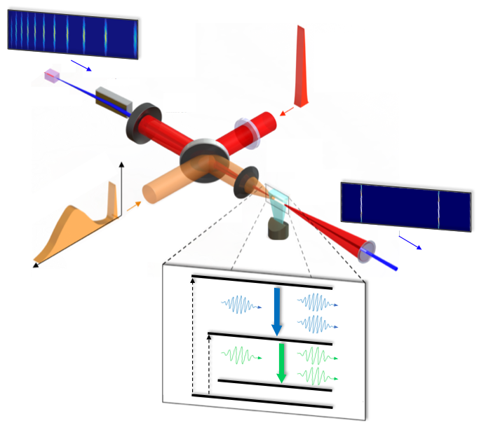
Two color soft xray laser Laboratoire d'Optique Appliquée
Our innovative color x ray imaging technology, visual software and computerized x-rays enable real-time, non-intrusive inspections from multiple viewing angles. Supported by a visionary team of experts, we push the limits of threat detection with solutions that achieve true security for communities around the globe. 6 COLOR IMAGING; 8 COLOR IMAGING

World's First 3D Color XRays of Human Body Produced Using CERN Technology
Some months back, CERN announced the first 3D color X-ray of a human made possible using the Medipix devices. The result is a high-resolution, 3D, color image of not just living structures like.
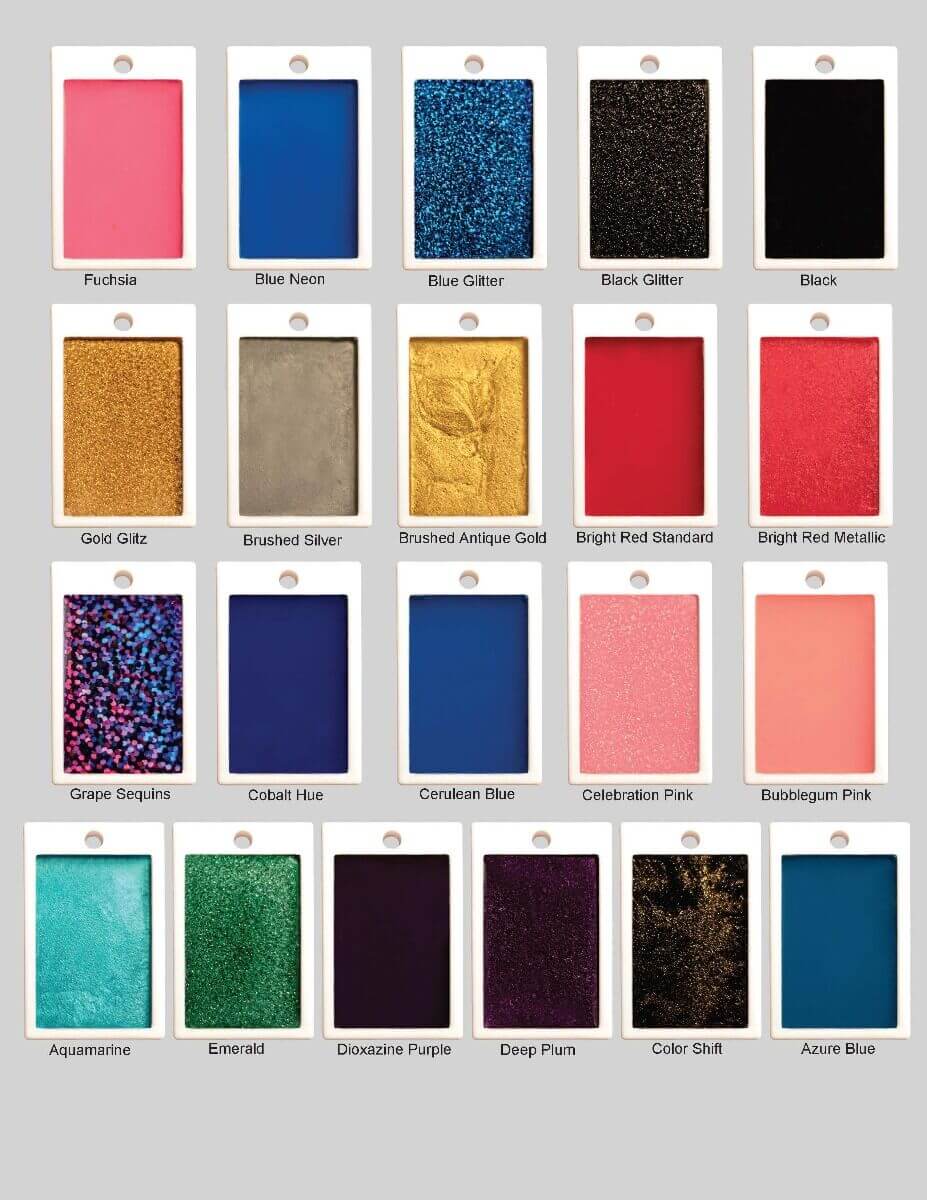
Plastic Lead X Ray Markers Mix & Match Glitter Color Magic Xray Markers
Colour x-ray is the future: an introduction to MARS 28 Mar 2021 Mars Bio Imaging User Uncategorized In early 2000, a young radiologist was posed this question over a beer: What does the word 'futuristic' mean to you?

Chandra 3Color Xray Image of N 63A ESA/Hubble
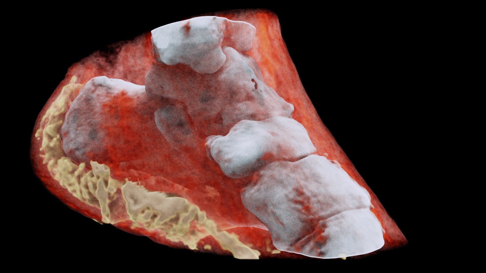
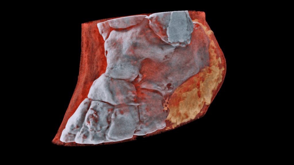
Stunning new color X-ray images, from a company called Mars Bioimaging, in New Zealand, seem to make flesh and bone translucent and hyperreal. A scan of an ankle rotates in this GIF. (Image.

Color Xray *CUP
49K 1.4M views 3 years ago At the University of Canterbury, in Christchurch, New Zealand, the team at Mars Bioimaging are using detector equipment originally developed for the Large Hadron.

18 best Chiropractic Color XRays and MRI of the Neck (Cervical Spine) images on Pinterest
What you can expect During the X-ray X-rays are performed at doctors' offices, dentists' offices, emergency rooms and hospitals — wherever an X-ray machine is available. The machine produces a safe level of radiation that passes through your body and records an image on a specialized plate. You can't feel an X-ray.

7 best Color MRI and XRays of Degenerative Disc Disease of the Neck images on Pinterest
The boring old black-and-white X-ray slides are a thing of the past — after 10 years spent in development, MARS Bioimaging has unveiled the first-ever color X-ray scanner. The device offers.

World’s First Full Color 3D XRays Technology To See Human Body
That means two objects of similar density but different materials can be distinguished using color x-ray, but not traditional x-ray. How does MARS make color images? In-house algorithms process the energy information from the x-ray to determine the materials present. Then, you can apply an arbitrary color (or color range) to each material.
A New CT Scanner Captures 3D ColorXRays
For their new method, the scientists used an X-ray color camera developed by PNSensor in Munich and a novel imaging system that essentially consists of a specially structured, gold-coated plate between the object and the detector, which means the sample casts a shadow.
10 Medical Advances that Sound Like Science Fiction
Specific densities are assigned different colors, so that bones appear white, muscle appears red, fat appears yellow, and implants can be blue or green. The models are not only incredibly striking,.
First human scanned with nextgeneration 3D color medical scanner Tech Explorist
What is color x-ray? Color x-ray is where the energy of the x-rays that pass through the object is measured. This technology is not a false color of an x-ray density map. The "color" in these images is actually true x-ray color and is not meant to represent the visible color of a material.

3D color Xray machine heads for trials
A 3D image of a wrist with a watch showing part of the finger bones in white and soft tissue in red. (Image: MARS Bioimaging Ltd) So far, researchers have been using a small version of the MARS scanner to study cancer, bone and joint health, and vascular diseases that cause heart attacks and strokes.
Color XRay imaging is just around the corner and we have the photos to prove it
Manchester University. "3-D color X-Ray imaging radically improved for identifying contraband, corrosion or cancer." ScienceDaily. www.sciencedaily.com / releases / 2013 / 01 / 130107082224.htm.
The Unofficial Guide to Radiology 100 Practice Chest Xrays
3-D Color X-Rays Could Help Spot Deadly Disease Without Surgery A new medical scanner, derived from technology used by particle physics researchers at CERN, "is like the upgrade from.

Medicine and Technology Future Color Xray technology illustrated
New Zealand scientists have performed the first-ever 3-D, colour X-ray on a human, using a technique that promises to improve the field of medical diagnostics, said Europe's CERN physics lab which.